The Radiographic Imaging Of Blood Vessels Of The Eye With Fluorescing Dye Is - Radiographic image of an artery: This process can examine retinal vessels in the case of diabetes.
Fluorescein Angiography - Retina-vitreous Surgeons Of Cny
Radiographic imaging of blood vessels (of the eye with fluorescent dye) term.

The radiographic imaging of blood vessels of the eye with fluorescing dye is. Blood flow exams often involve fluoroscopy. Diagnostic terms built from word parts 52. Fluoroscopies are used to evaluate both hard and soft tissue, including bones, joints, organs and vessels.
Common imaging types include ct (computer tomography), mri (magnetic resonance imaging, ultrasound and. And the ultrawide imaging captures pictures of structures on the periphery of the eye. Abnormality here could indicate head injury or damage to the brain)
Histamine binds to specific receptor sites. A term meaning condition of equal pupil (size) is. Radiographic image of a vein:
Instrument used for visual examination of the eye. Radiographic imaging of blood vessels(of the eye with fourescing dye) keratometer: Radiographic imaging of blood vessels (of the eye w/ fluorescing dye) keratometer instrument used to measure the curvature of the cornea for fitting contact lenses
The instrument used for visual examination of (the interior) the eye is the ophthalmoscope the radiographic imaging of blood vessels (of the eye with fluorescing dye) is Visual exam of the eye… Instrument used to view vessel:
Instrument used for visual exam of the interior of the eye: Scanning lung visual imaging of the distribution of ventilation or blood flow in the lungs by scanning the lungs after the patient has been injected with or has inhaled radioactive material Radiology may be divided into two different areas, diagnostic radiology and interventional radiology.
Which condition involves leakage of blood and other fluids from intraocular vessels that destroy vision cells, leading to permanent vision loss? This makes it easier to spot any abnormalities. Blood vessels (yellow) and lymphatic vessels (blue) can be visualized in the same tissue with appropriate processing (c) (vakoc et al., 2009).
Instrument used to measure the cornea (used for fitting contact lenses) ophthalmoscope: Lymphatic vessels (green) and blood vessels at day 1 (d), day 8 (e), day 15 (f) and day 22. The normally expected flow void of the left carotid artery is absent (arrow).
Record or image of a blood vessel term. The radiographic imaging of blood vessels (of the eye with fluorescing dye) is a. Radiology is a branch of medicine that uses imaging technology to diagnose and treat disease.
Record of heart using sound: Radiographic imaging of blood vessels (of the eye with fluorescing dye) keratometer. Medical imaging remains one of the best ways to diagnose patients, as it allows us to see what’s going on inside the body without the need for surgery or other invasive procedures.
Radio imaging of blood vessels: Radiographic imaging of blood vessels (of the eye with fluorescing dye). Vis examination of blood vessel:
In these procedures the animal is given a dye that will block x‑rays. This can be given intravenously to examine organs like the kidneys or heart, or by mouth to examine the digestive tract. Rad imaging of a vein:
Doctors who specialize in radiology are called radiologists. Radiographic imaging of blood vessels (of the eye with fluorescing dye) keratometer instrument used to measure (the curvature of) the cornea (used for fitting contact lenses) A diagnostic procedure in which a narrow beam of light is focused onto parts of the eye to permit the doctor to examine the structure at the front of the eye perrla pupils are equal, round, response to light, and accommodation;
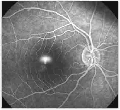
Instrument used to measure the cornea. Final not built test and treatment. Fluorescein angiography is the medical term meaning radiographic study of the blood vessels of the retina.
H1 receptors are found in endothelial The procedure is often done with contrast dyes, which show how they flow through the body. “the impact of integrating all three systems into one piece of equipment is significant.” the impact of integrating all three systems into one
Principal locations are the skin, respiratory tract, gi tract, and blood vessels. Instrument used to measure (the curvature of) the cornea (used for fitting contact lenses) term. Blood test used to measure the level of creatine phosphokinase, an enzyme of heart and skeletal muscle released into the blood after muscle injury or necrosis.
A test to determine the amount of glucose (sugar) in the blood after fasting for 8 to 10 hours. Radiographic imaging of blood or lymph vessels which have been injected with dye;
Choroidal Osteoma New York Eye Cancer Center

Moran Core Fluorescein

Pdf Magnetic Resonance Angiography Source Images In Carotid Cavernous Fistulas

Fluorescein Angiography Eye Physicians And Surgeons Of Ontario

Pdf Imaging Review Of Ocular And Optic Nerve Trauma

Pdf Imaging Review Of Ocular And Optic Nerve Trauma
Diagnostics Free Full-text Classification Of Non-infectious Andor Immune Mediated Choroiditis A Brief Overview Of The Essentials Html

Fluorescein Angiography - Wikipedia
Choroidal Hemangioma New York Eye Cancer Center
Fluorescein Angiography Department Of Ophthalmology
Fluorescein Angiography

Imaging For Neuroophthalmic And Orbital Disease A Review - Lee - 2009 - Clinical Experimental Ophthalmology - Wiley Online Library

Anatomy Of The Optic Nerve And Visual Pathway - Sciencedirect
Fluorescein Angiography - Retina-vitreous Surgeons Of Cny
